Oldham Lab Overview
3D and 4D Radiation Dosimetry
We have developed a range of uniquely capable state-of-the-art 3D dosimetry systems with funding support from the National Institute of Health. These systems are currently being applied to a diverse range of challenges in both the clinical (radiation therapy) and research domains.
Optical-CT/ECT Imaging
The lab has pioneered novel imaging techniques of optical-computed-tomography (optical-CT), and optical-emission-computed-tomography (optical-ECT). These techniques have the potential to provide uniquely useful information on biological processes in bulk unsectioned tumor and tissue samples. Click here for more information.
Radiation and Immunotherapy
A major recent focus has been several collaborations investigating new ways to use radiation to help activate a long term immune response. Click here for more information.
3D Printing and Pre-Clinical Micro-IMRT
A collaboration with The Kirsch Lab focuses on implementing a micro-IMRT capability for pre-clinical work utilizing advanced 3D printing techniques.
Featured Articles in the News
Cherenkov photo-activation enhances radiotherapy (2018)
3D Dosimetry Protocol is Compatible with MR-Guided Radiotherapy (2017)
Radiotherapy Scheme Spares Hippocampus in Rats (2017)
3D printed dosimeters aid SBRT research (2015)
Assessing micro-irradiator accuracy (2013)
Optical ECT: the complete correction (2010)
Optical-ECT: a quantitative approach (2008)
Seeing tumours in a new light (2007)
Research
The lab pursues three main avenues of research as described below.
Radiation and Immunotherapy
For the last five years, we have actively pursued a new research direction involving radiation and immunotherapy. This research uses radiation to photo-activate bio-therapeutics in deep tissue with the potential to stimulate long-term anti-tumor immunity. Two novel and patented treatment paradigms are introduced: X-ray Photo-Activated Cancer Therapy (X-PACT) [1], and Radiotherapy Enhanced by Cherenkov photo-Activation (RECA) [2]. These papers both won the SEAAPM Best Paper in Medical Physics Award for the years 2017 and 2018 respectively. Dr. Oldham was the PI on a Duke Brain Spore development project program grant to develop X-PACT.
[1]: Yoon, S.W., V. Tsvankin, Z. Shrock, B. Meng, X. Zhang, M. Dewhirst, P. Fecci, J. Adamson, and M. Oldham, Enhancing Radiation Therapy Through Cherenkov Light-Activated Phototherapy. Int J Radiat Oncol Biol Phys, 2018. 100(3): p. 794-801
[2]: Oldham, M., P. Yoon, Z. Fathi, W.F. Beyer, J. Adamson, L. Liu, D. Alcorta, W. Xia, T. Osada, C. Liu, X.Y. Yang, R.D. Dodd, J.E. Herndon, 2nd, B. Meng, D.G. Kirsch, H.K. Lyerly, M.W. Dewhirst, P. Fecci, H. Walder, and N.L. Spector, X-Ray Psoralen Activated Cancer Therapy (X-PACT). PLoS One, 2016. 11(9): p. e0162078
High Resolution 3D Dosimetry
Founded in 2005, Duke 3D Dosimetry Lab has established itself as one of the leading 3D dosimetry groups in the world. The Lab has received NIH R01 funding to develop optical imaging techniques for 3D dosimetry. Key aims of the Lab include elevating the state-of-the-art for verification of advanced radiation therapy treatments, including radiosurgery, VMAT, deformable dose tracking, proton-therapy, micro-beam therapy, pre-clinical cancer research and the nuclear industry. Our research has demonstrated that the various 3D dosimetry system can achieve these aims and has demonstrated many firsts. The lab has won the AAPM Farrington Daniels Award for the best dosimetry paper in the journal of Medical Physics in 2001 [3] and 2015 [4].
[3] Oldham, M., J.H. Siewerdsen, A. Shetty, and D.A. Jaffray, High resolution gel-dosimetry by optical-CT and MR scanning. Med Phys, 2001. 28(7): p. 1436-45
[4] Bache, S.T., T. Juang, M.D. Belley, B.F. Koontz, J. Adamovics, T.T. Yoshizumi, D.G. Kirsch, and M. Oldham, Investigating the accuracy of microstereotactic-body-radiotherapy utilizing anatomically accurate 3D printed rodent-morphic dosimeters. Med Phys, 2015. 42(2): p. 846-55
Hi-Res Optical 3D Bio-Imaging of Cleared Tissue
The Lab has a long-term collaboration with Dr. Mark Dewhirst’s group at Duke, which is focused on novel techniques for optical bio-imaging – specifically, applying optical tomographic techniques to imaging un-sectioned tissue samples. The primary aim is to investigate and optimize the new techniques of optical-computed-tomography (optical-CT) and optical-emission-tomography (optical-ECT), and establish their capability as powerful new tools of discovery in pre-clinical cancer biology. Optical-CT/ECT are the optical analogues of x-ray-CT and SPECT respectively. In conjunction with new techniques for rendering bulk tissue samples transparent to visible light, optical-CT/ECT can yield unique data unattainable by other modalities. Principally: high-spatial resolution (potentially 10-20µm), high contrast, precisely co-registered, three-dimensional (3D) images of the distribution of fluorescent reporter proteins and absorbing contrast agents in large un-sectioned tissue samples (up to ~8cc). This capability is of significant interest to cancer researchers because it provides, for the first time, data similar to that obtained from influential window-chamber studies, but in much larger tumors. Optical-CT/ECT have potential for very diverse application. Our initial work aims to image the 3D distribution of vasculature, HIF-1 and viable tumor in much larger tumors than has been possible before. Two papers from this research collaboration were selected for Phys Med Biol Highlights Edition in 2008 [5] and 2010 [6], and one was short-listed for the Robert’s Prize from the IOP.
[5] Oldham, M., H. Sakhalkar, T. Oliver, G. Allan Johnson, and M. Dewhirst, Optical clearing of unsectioned specimens for three-dimensional imaging via optical transmission and emission tomography. J Biomed Opt, 2008. 13(2): p. 021113
[6] Thomas, A., J. Bowsher, J. Roper, T. Oliver, M. Dewhirst, and M. Oldham, A comprehensive method for optical-emission computed tomography. Physics in Medicine and Biology, 2010. 55(14): p. 3947-3957
Publications
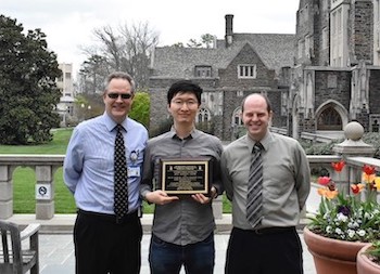
In 2018, the Lab won the SEAAPM Best Paper Award for our paper entitled “Enhancing Radiation Therapy Through Cherenkov Light-Activated Phototherapy."
In the picture, Suk-Whan (Paul) Yoon (first author) is holding the award, with advisors M. Oldham (left) and Justus Adamson (right).
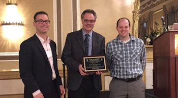
In 2017, the lab won the SEAAPM Best Paper Award for our paper entitled “X-Ray Psoralen Activated Cancer Therapy (X-PACT).”
Oldham M, Yoon P, Fathi Z, Beyer WF, Adamson J, Liu L, Alcorta D, Xia W, Osada T, Liu C, Yang XY, Dodd RD, Herndon JE 2nd, Meng B, Kirsch DG, Lyerly HK, Dewhirst MW, Fecci P, Walder H, Spector NL. Click here to view it.
Picture taken at the Awards Ceremony at the 2017 Annual SEAAPM meeting in Charleston, SC. Left to right, David Wiant, PhD, (Past president), M. Oldham and J. Adamson.
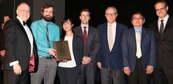
The 2016 Awards Ceremony at the AAPM Annual Meeting in Washington, DC. Left: Steve Bache, MS (first author) and Matt Belley, PhD. Right: Bruce Currant (AAPM President), Steve Bache, Titania Juang, Matt Belley, John Adamovics (Heuris), Terry Yoshizumi and Mark Oldham.
The Farrington Daniels Award is given every year for the best paper published in Medical Physics on Radiation Dosimetry. Click here to view it.
View Dr. Oldham's NIH Bibliography page or Google Scholar page.
People

Resources
The Duke 3D Dosimetry Lab has received NIH R01 funding to develop optical imaging techniques for 3D dosimetry. Our research has demonstrated that a new PRESAGE/optical-CT system is a viable 3D dosimetry system for radiation therapy and general radiation measurement. Our research involves a strong 3-party consortium to pursue two major aims; a continued technical and investigational aim, and a new clinical applications aim. The consortium combines outstanding expertise in 3D dosimetry (Duke) with credentialing experience (Geoff Ibbott at the RPC), and materials expertise (John Adamovics at Rider University). The aims balance investigating innovations to facilitate 3D dosimetry for the non-specialized clinic, while still addressing the urgent need for detailed clinical IMRT dosimetry studies using a more specialized system.
We hypothesize novel optical-CT scanning systems, and novel radiochromic formulations will achieve substantial increase in speed and practicality without compromising accuracy. We also hypothesize that 3D dosimetry techniques will detect more clinical IMRT deliveries failing standard comparison metrics, than conventional 2D techniques. This research will conduct the first comprehensive 3D investigations of the accuracy of IMRT deliveries in cohorts of patients in key clinical sites (pelvic, head-and-neck, and thorax). Successful completion will elevate the state-of-the-art for verification of advanced radiation treatments, and may demonstrate the feasibility of a powerful and comprehensive new 3D credentialing technology with potential to elevate the standard of practice in clinical trials.
The lab has access to ~600sq feet of lab-space in the department of Radiation Therapy Physics. This space includes a dedicated imaging room with dark-room facilities, and a fume hood. The lab contains several unique optical-CT scanning systems, developed in-house, for various applications in 3D dosimetry and biological imaging. We are open to collaborations with other researchers interested in 3D dosimetry, or high-resolution 3D imaging of fluorescent tissue markers.
Contact
Director, Duke Medical Physics Graduate Program
Duke University Medical Center
Durham, NC
919-668-0349
Email Dr. Oldham at mark.oldham@duke.edu